 When talking about organelle size, most people believe the bigger the cell, the larger the organelle should be. In fact, this is not true, at least for mitotic spindles. Recent studies showed that the mitotic spindle size scales with the cell size in small cells, but approaches an upper limit in large cells. However, how the spindle size is sensed and regulated still eludes scientists.
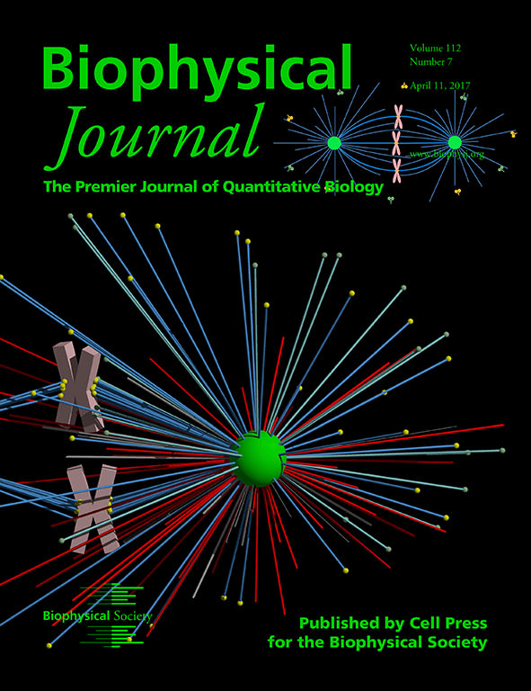
The cover image for the April 11 issue of the Biophysical Journal shows a configuration sampled by a three-dimensional computational simulation guided by a general model for mitotic spindles. The model explicitly shows microtubules (colorful rods), centrosomes (green sphere), and chromosomes (pink bulks). Microtubules can be nucleated from the centrosomes, grow outward, and show the dynamic instability. When microtubules encounter the cortex or chromosome arms, they can generate pushing forces (red rods) due to the polymerization of microtubules. Various molecular motors on cortex and chromosomes, including dyneins (yellow dots) and kinesins (green dots), can bind to microtubules and generate pushing forces (green rods) or pulling forces (blue rods). Therefore, the centrosomes and chromosomes can move under these forces. In this way, the mitotic spindle can be self-assembled to form a bipolar structure with certain size, positioned to the cell center, and orientated to the long axis. Meanwhile, the chromosomes can be attached correctly and aligned on the equatorial plate.
This computational model is very useful for studying the size regulation of mitotic spindles. The spindle size is usually defined as the pole-to-pole distance, so that the problem of spindle size regulation is dependent on the positioning of two poles. The position of each pole is determined by the mechanical equilibrium between the cortical force and the chromosome force on the spindle pole. For each pole, the chromosomes and the cortex are geometrically asymmetric. In small cells, the geometric asymmetry is small and the pole is nearly positioned to at the center of each half cell so that the spindle size scales with the cell size. However, in large cells, because few microtubules can reach the cell boundary, the geometric asymmetry is large and the spindle size is only determined by the chromosomes; that is, the spindle size approaches the upper limit. Therefore, this work revealed a novel and essential physical mechanism of the spindle size regulation.
There are certainly many other factors that influence spindle size but only quantitatively; they cannot explain the existence of the size limit of spindles.
This computational model provides a very powerful and robust tool. It can be combined with existing biochemical techniques to explore many important and interesting phenomena, including the positioning and orientation of mitotic spindles, the spontaneous oscillation of chromosomes, the mechanical response of spindles under various forces, and many other relevant questions.
- Jingchen Li and Hongyuan Jiang