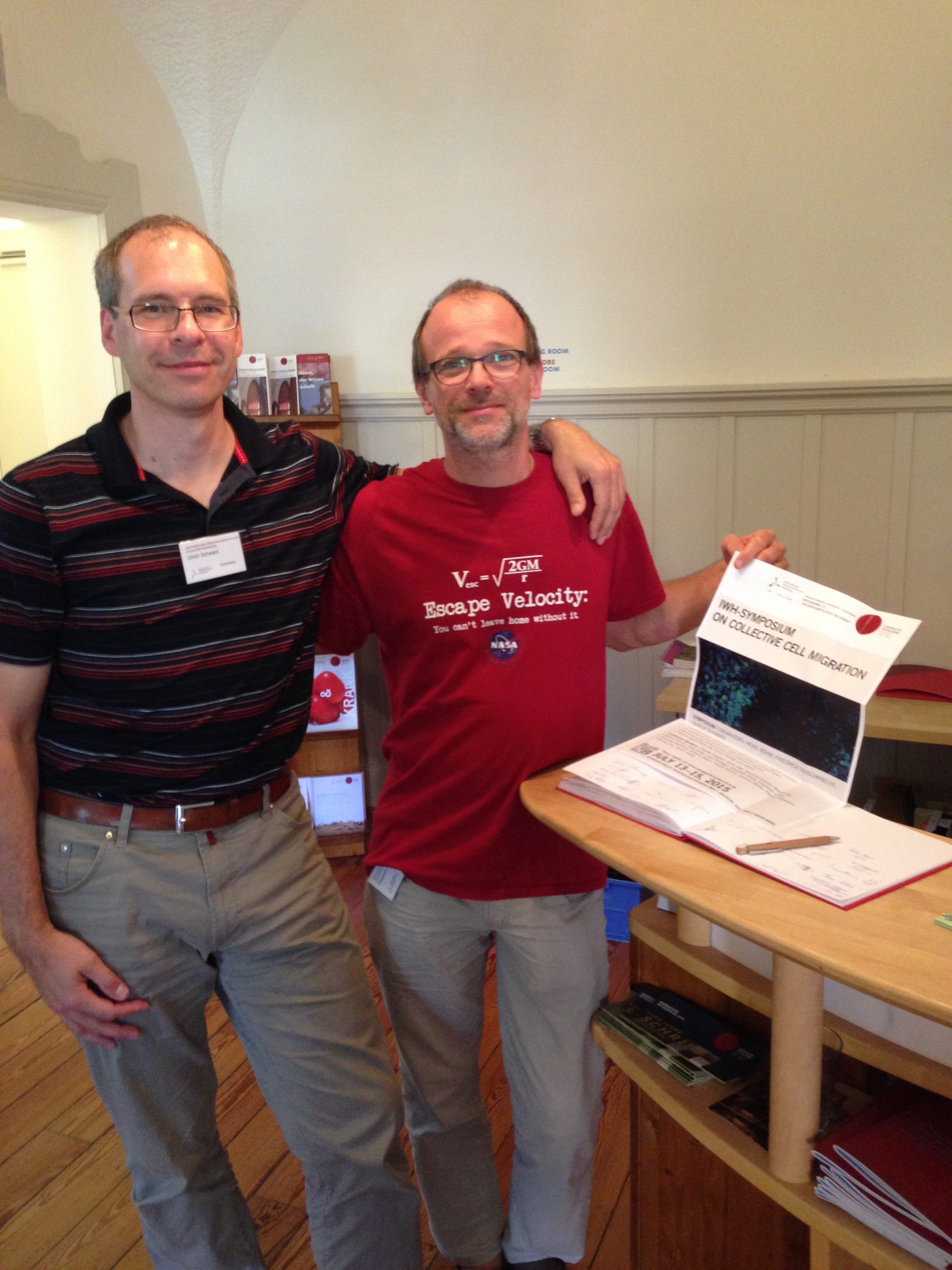
April 25 is World Malaria Day. Malaria is an infectious disease that is transmitted by mosquitoes and takes around half a million lives every year. It is caused by unicellular parasites from the genus Plasmodium that have left a bigger imprint on our genome than any other parasite to date. In recognition of World Malaria Day, we spoke with BPS member Ulrich Schwarz from the Department of Physics and Astronomy at Heidelberg University and his collaborator Friedrich Frischknecht from Heidelberg University Medical School about their research relating to malaria.

What is the connection between your research and malaria?
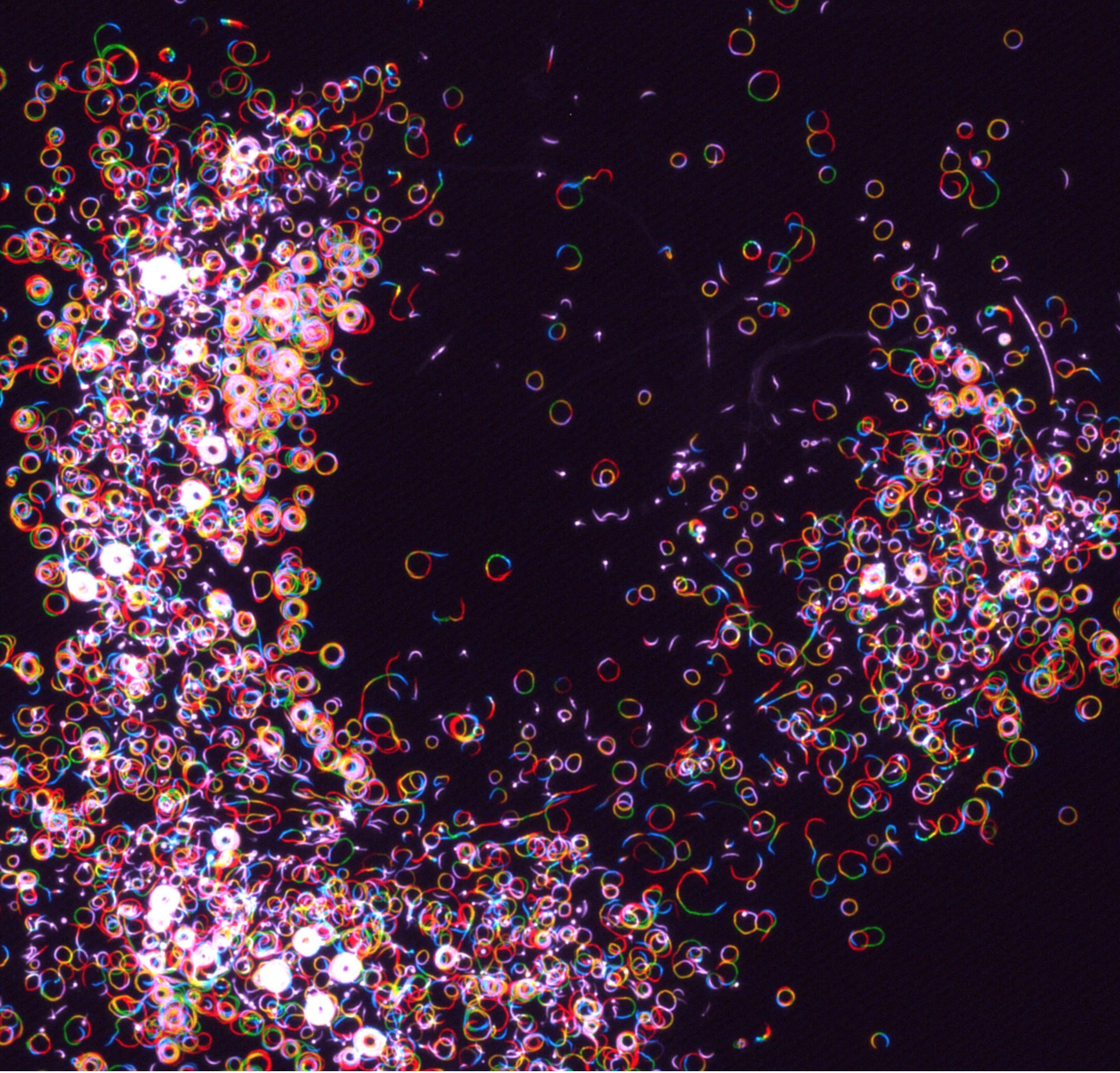
We both started out with an interest in the actin cytoskeleton, which is a major determinant of mechanics, adhesion and migration of human cells. It is also essential for many cellular processes used by the malaria parasite as it makes its journey through the human host. For example, after being released by a mosquito bite, the parasite (in a torpedo-like form called the “sporozoite”) rapidly moves through the host skin with a velocity of 1-3 um/s that is unsurpassed by any other cell type moving in an adhesion-dependent manner. This surprising achievement is based on “gliding motility,” where a thin layer of parasite actin is pushed from the front to the back of the parasite by myosin motors. Once this layer connects to the environment through adhesion molecules, the parasite itself is pushed forward. However, how the different parts of this system work together and how sporozoites manage to achieve such high velocities is still a riddle.
Another fascinating example is how P. falciparum, the most virulent variant of the malaria-causing parasites, uses the actin cytoskeleton of the host cell: once inside red blood cells it starts to mine actin from the spectrin-actin network of the red blood cell to build a unique transport system that brings parasite-made proteins to the surface of the host cell, including adhesion molecules. The infected red blood cells become sticky and thus increase its residency time in the vasculature, thereby avoiding clearance by the spleen. It is still unknown how the parasite manages to coordinate all the different changes at the red blood cell surface from a distance and how this exploits the actin dynamics of the host cell. We believe that these and other open questions in malaria research can only be completely answered by biophysical approaches, combining quantitative experiments with mathematical models.
Why is your research important to those concerned about malaria?
Malaria is a devastating disease and there is huge medical interest in any potential new therapy to fight it. However, efforts in this direction are often focused on genetic and biochemical ways to interfere with the malaria life cycle, which despite its complex nature seems to offer relatively few weak spots to intervene. We believe that biophysical approaches might open up new avenues to think about this disease and to fight it. For example, a successful infection seems to depend on a single sporozoite making its way from the skin to the liver, where it then can multiply into tens of thousands of merozoites and finally invade red blood cells. If we were able to interfere with the actomyosin machinery during sporozoite gliding motility in the skin or merozoite invasion of red blood cells, we might have found new inroads for a therapy. Also, the parasite-induced actin-based transport system in the infected red blood cells seems to be a critical element for disease progression and any biophysical insight into its function might help to find a new point of attack. In the permanent struggle between mankind and its pathogens, biophysics might soon play an equally important role as genetics and biochemistry already do. Finally one should note that malaria is not only a devastating disease, but also a fascinating research subject, from which we can learn a lot, e.g. about evolution and function of the actin cytoskeleton, and that our interdisciplinary approach allows training of students from different disciplines in malaria research.
How did you get into this area of research?
A bit more than 10 years ago, we both worked as junior research group leaders at Heidelberg, where today we both work as professors. Ulrich Schwarz worked on theoretical physics approaches to cell adhesion and mechanics, and Friedrich Frischknecht worked on imaging sporozoite motility. We bumped into each other at seminars and quickly realized the synergy that can be achieved by quantitatively measuring the forces that sporozoites exert on their environment during gliding motility, using traction force microscopy on soft elastic substrates, which at that time was still a relatively novel method in cellular biophysics. Later we joined forces with Joachim Spatz, from the Institute for Physical Chemistry at Heidelberg University, and extended the range of biophysical methods to also include reflection interference contrast microscopy, pillar assays, optical tweezers and biofunctionalized surfaces. In our joint work, we put large emphasis on quantification, because we think that real understanding is only achieved when the numbers add up.
How long have you been working on it?
Our first paper including traction force microscopy with sporozoites came out in Cell Host Microbe in 2009, and since then we collaborate on a continuous basis with each other and in an increasingly larger group, including colleagues from physics, physical chemistry, biology and medicine.
Do you receive public funding for this work? If so, from what agency?
We started off with the support by our national junior research group grants and later got very helpful support through the German excellence initiative, both by Heidelberg University intramural funding and the cluster of excellence CellNetworks. Our collaboration also helped Friedrich Frischknecht obtaining a starting grant from the European Research Council (ERC) in 2011. Since 2014, we are also funded by the German Research Foundation (DFG) through the Collaborative Research Center 1129 (www.sfb1129.de) on quantitative analysis of pathogen replication and spread, in which currently 4 out of the 15 projects are on malaria. We are very happy that here at Heidelberg, interdisciplinary projects on the biophysics of malaria have found generous support from the very beginning, and in general we hope that the relation between physics and parasitology will gain more traction in the future. In contrast to some other countries, the DFG funds basic research without a necessity for immediate translational potential, so we can focus on the fundamental biophysics questions raised by malaria research.
Have you had any surprise findings thus far?
Plenty. To start with, Friedrich Frischknecht was part of a team to show that the skin stage is an important and formerly somehow overlooked part of the malaria lifecycle, because earlier it was believed that the mosquitoes inject the parasites directly into the human blood vessels. Motility was assumed to be a smooth process as the parasites do not change their shape as they move. In our work with traction force microscopy, we then discovered that gliding motility is not a continuous process at all, but consists of a sequence of stick-slip events. Using pillar assays and in vivo imaging, we then were able to demonstrate that the geometry of the environment determines how gliding motility plays out, as also confirmed by an agent-based mathematical model.
Our most surprising finding from the physics point of view was that sporozoites can engage in collective cell migration. Usually studied in the context of development, wound healing or cancer cell migration, we found that collective cell migration is also crucial for sporozoites to exit from the oocytes, where the parasites form at the wall of the mosquito gut, and likely also within the salivary glands, where the parasites wait for injection. In 2015, we even organized a meeting on collective cell migration at Heidelberg (with financial support from the excellence initiative for workshops in the area of scientific computing), to exchange ideas with other biophysicists working on collective cell migration.
What is particularly interesting about the work from the perspective of other researchers?
Each winter term we jointly offer a seminar on cell migration for physics and biology students. If it comes to malaria, we always make the point that although gliding motility is based on the same physical principles as cell migration of more familiar model systems also covered in the seminar (e.g. keratocytes, fibroblasts and neutrophils), in detail it is very different. In the malaria parasite, actin filaments polymerize like in the other cellular systems, but the actin filaments tend to be short, highly dynamic and they do not branch. Like in the other model cellular systems, myosin leads to retrograde flow, but the exact spatial coordination and nature of the flow is unclear. If it comes to the actin-based transport system induced in the infected red blood cells, surprisingly one now can find branching, possibly because here the parasite uses host actin. Thus the malaria parasite teaches us a lot about the common principles and distinct differences between different biological systems, and one should always be open for surprises, in the sense that the malaria parasite has evolved different mechanisms than found in human cells.
What is particularly interesting about the work from the perspective of the public?
Malaria research has very high biomedical relevance, due to the large number of deaths every year and the huge economic burden it puts on the affected countries, which are located mainly in the equatorial regions. Of course it is gratifying to think that fundamental biophysics research contributes to the global effort in fighting this disease, but one also should note that many of the crucial issues in the medical context are not purely scientific, like prevention measures (e.g. mosquito nets) or accessibility to drugs. Working on malaria always creates attention in the public, but as a scientist, one should be careful to explain what can be expected from fundamental research and what not. In outreach events, we usual experience that the public is very much interested in imaging the amazing journey that the parasite takes through the bodies of its human and mosquito hosts.